

Post by: Dr. Anoop Kumar Singh
in Neurosurgery
Dr.Anoop Kumar Singh, Case Study
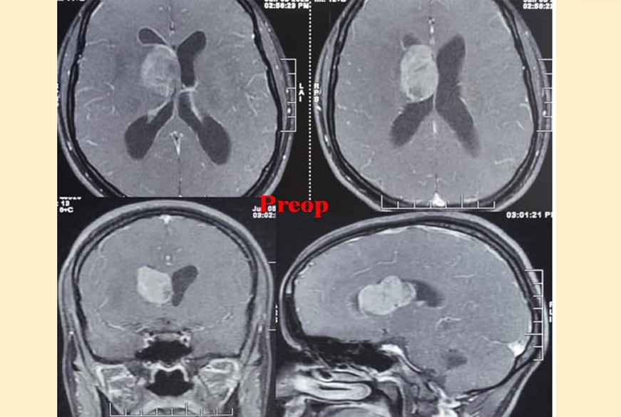
24 Yrs/F, H/O headache & vomiting for 15 days, O/E no neurological deficit, MRI brain showed Rt BG contrast-enhancing lesion. She underwent a conventional transcallosal interhemispheric approach and by creating corridors on either side of the Rt pericallosal artery, I resected the lesion. She had developed an evolving communicating hydrocephalus in postop, for which a VPMP shunt was done.
The biopsy was ependymoma. Finally, a neurologically intact patient was discharged on the 14th day after the first surgery.